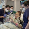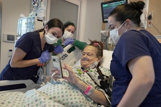
Scientists from Tomsk Polytechnic University are creating 3D-printed models of children's hearts. They are printed based on magnetic resonance imaging (MRI) of real patients. These models are used as simulators for cardiac surgeons to plan and pre-work forthcoming operations.
The new project for 3D-printing human hearts is developed on the basis of the TPU Modern Manufacturing Technology Scientific and Educational Center. The university scientists carry out it for the Tomsk Research Institute of Cardiology (RIC). The center fellows print plastic copies of real hearts on the basis of magnetic resonance imaging. Having a tangible model, cardiac surgeons will be able to study organ defects in detail and choose proper surgical treatment plan.
The first 3D-models of adult hearts were printed at TPU in summer. Now TPU scientists start 3D printing of children's hearts.
Tomsk cardiologists have very different patients: from elderly people who have lived to adulthood with childhood pathology to very infants who were diagnosed a rare heart disease. According to doctors, operating children is twice difficult since children's hearts are several times smaller than adults' ones. Therefore, all actions of surgeons should be thought out in advance, accurate and adjusted.
Professor Vyacheslav Ryabov from the SSMU Department of Cardiology, Deputy Director for Research and Clinical Care at RIC, Head of Emergency Cardiology Department says, "Our cardiac surgeons suggested creating 3D-models of hearts. Today surgeons do not blindly go to a planned operation. Using modern diagnostic visualization technologies they try to obtain information prior operations, which can help to plan operation process."
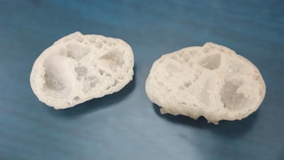
Now, 3D-simulation is traditionally used in these cases. According to him, a disadvantage of computer models is in presenting only external surface of hearts, internal surfaces are available only at single sections. 3D-models are possible to open as pyramids and to look what anomalies are located inside the heart.
Leading research fellow at the RIC Laboratory of Radionuclide Testing Methods, doctor of medical sciences Konstantin Zavadskiy, says "For example, when we receive patients with rare pathologies, we always want to get 3D-models and see how these pathologies are actually arranged. Moreover, medical residents and PhD students study at the institute and thanks to these simulators they will be able to investigate rare and complex interrelations of anomaly structures inside the hearts of sick people that will help them to become good physicians."
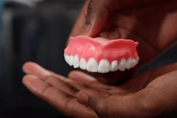
Currently, the scientists print 3D-models of children's and adult's hearts out of plastic. Further, they plan to print heart copies out of the rubber-like material which structure is similar to natural heart tissue.
Such training will bring surgeons as close as possible to real conditions. Another goal of TPU scientists is to shorten time of heart 3D printing.
Director of the Modern Manufacturing Technology Scientific and Educational Center Vasily Fyodorov summarizes, "We need two weeks to create a heart 3D-model. Meanwhile, patients often need urgent help. We should reduce printing time to 1-2 days. For this purpose, it is required to select an appropriate equipment and to develop proper software that will directly convert MRI data into the stl files for 3D printing. Now we are looking for funding to develop software. Ideally, we would like the 3D printer to be in the hospital."